The Veterinary Dental Center of Atlanta is pleased to announce that our team of experienced veterinarians has successfully diagnosed and treated a dentigerous cyst in a beloved Boston Terrier dog. The diagnosis was made using full mouth radiographs, which provided a clear and comprehensive view of the dog’s dental condition.
Our team then discussed a treatment plan with the pet owner, taking into consideration the dog’s overall health and well-being. In veterinary dentistry, it is not uncommon to encounter pathology that is more significant than what appears on the surface. This was the case with the Boston Terrier dog in question, but our team was able to provide the necessary care and treatment to ensure a positive outcome.
A Dentigerous Cyst in a Boston Terrier dog is diagnosed with full mouth radiographs and a treatment plan is discussed. In veterinary dentistry we commonly see pathology that is more significant that what appears of the surface. That was the case here.
This is the surprise case of the week. We’ve got a case that we saw this week and we’re going to show you some images of that and ask you what you think so here’s first image. This is a little Boston Terrier, 9 years old, that presented for tooth fracture. What we found with full mouth radiographs was much more significant than a tooth fracture.
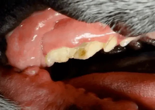
This is the tooth after we’ve kind of manipulated it a little bit, that whole back segment of that right upper fourth premolar in this little Boston Terrier was totally fractured and the tooth was actually in half under the gum as well.
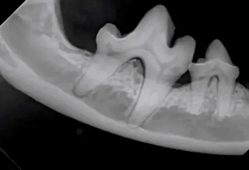
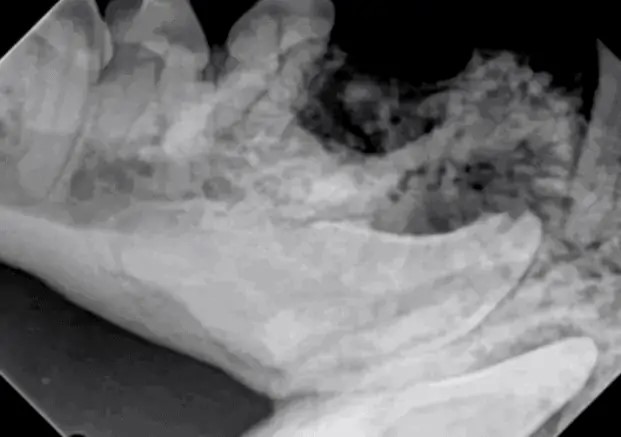
This is the rostral mandible on the right side. Anything unusual?
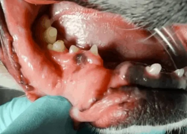
There’s the next image, there is a hole there and in that hole is a little foreign body. That might give you a good idea about what we’re dealing with. I’m looking into the chat here in the comments and Patricia, glad to have you again. Patricia’s from Brazil, she’s one of our friends over there. No canine tooth and that’s absolutely right, there is no canine tooth there and there’s also no first premolar. There is a cyst there and many times when we have teeth that do not erupt they form dentigerous cysts.
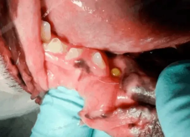
In this case, that is the case. We removed that little foreign body and that little foreign body turned out to be what I think is a pea, a little field pea that this dog evidently ate and it got stuck down in the tissue. Once I took that out, I just put some pressure on it and just exploded like a pea would although, it was a little bit yellowish. I think it had been there for a long time in that defect and caused all that mucositis there.
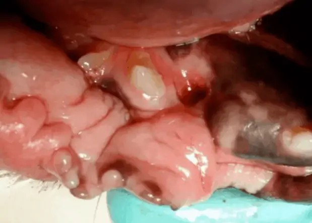
Pretty interesting but, here’s the image of that side and you can see that canine tooth is indeed unerupted. There looks like there is a remnant of the first premolar there as well in that dense area right at the bone interface above that canine tooth. Then you have all that bone proliferation.
There’s the other side. You can definitely see the remnant of that first premolar superimposed over that lucency there on the rostral lucency on the left of your screen. Again, you’ve got that canine tooth there, there looks like there’s a lucency mainly derived from that first premolar but, when we’re considering this case we have to take into consideration a couple things.
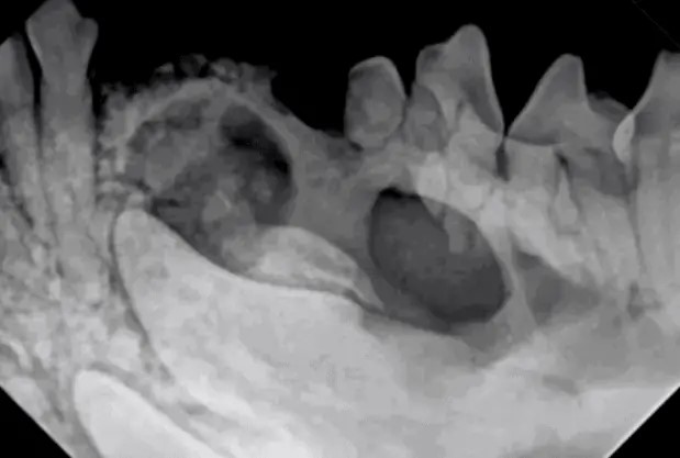
One is that this may, because his dog is so old, either of these may have undergone some metaplastic changes that could be neoplasia. We can get metaplastic changes that turn dentigerous cysts to neoplastic conditions. We definitely would want to biopsy that. Part of the approach would probably be initially to biopsy, remove as much of the cyst as we can, come back and finish later assuming that we might have neoplasia there and don’t want to disrupt all that tissue and spend all that time if mandibulectomy is going to be the way that we’re going to proceed rather than a cyst excision. Because of the patients age, because the cysts are not horrendous, we would probably biopsy both sides first, remove as much as we could, and then come back later to remove those canine teeth if indeed the biopsy is negative.

Update on this case. Biopsies confirmed dentigerous cyst with no evidence of neoplasia. Complete cyst excision and canine extractions were performed.
