Dental malocclusion, also known as an improper bite, is a common condition in dogs that can lead to a variety of dental and oral health issues. At the Veterinary Dental Center of Atlanta, we understand the importance of early detection and treatment of dental malocclusion in dogs.
This case study will provide an in-depth look at a patient at our center who was diagnosed with dental malocclusion and the treatment plan that was implemented to improve their dental health. We will also discuss the causes, symptoms, and potential complications of dental malocclusion in dogs, as well as the importance of regular dental check-ups and preventative care.
Identifying and preventing problems
Dogs deserve the same straight, engaging smile we humans seek, right? OK, that may be stretching it a bit, but what our patients do deserve is a bite that’s comfortable. This article highlights the classes of malocclusion. Future articles in this series will address individual cases that fall into these classes and show what can be done using both common and uncommon techniques to resolve problems.
Although preventing permanent teeth malocclusions is ideal, most of the cases veterinary dentists encounter consist of traumatic tooth-on-tooth or tooth-on-tissue disease in an adult patient that might otherwise have been prevented. Many patients present with severe tooth wear, fractures, endodontic disease and trauma to the palatal and lingual floor soft tissue or bone. So knowing normal anatomy is the first step in recognizing abnormal occlusions that can progress to more serious disease.
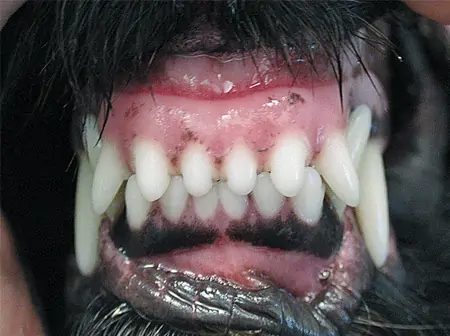
Photo 1: Normal occlusion of the incisors in a dog.
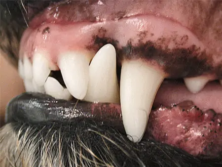
Photo 2: Normal occlusion of the canines in a dog.
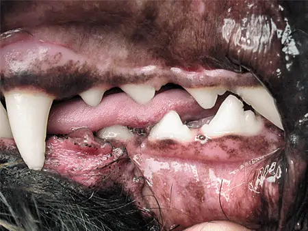
Photo 3: Normal occlusion of the premolars in a dog.
Types of Malocclusions
The American Veterinary Dental College (AVDC; www.avdc.org) describes dental malocclusions as follows:
- Distoversion: A tooth that’s in its anatomically correct position in the dental arch but is abnormally angled in a distal direction.
- Mesioversion: A tooth that’s in its anatomically correct position in the dental arch but is abnormally angled in a mesial direction.
- Linguoversion: A tooth that’s in its anatomically correct position in the dental arch but is abnormally angled in a lingual direction.
- Labioversion: An incisor or canine tooth that’s in its anatomically correct position in the dental arch but abnormally angled in a labial direction.
- Buccoversion: A premolar or molar that’s in its anatomically correct position in the dental arch but is abnormally angled in a buccal direction.
- Crossbite: A malocclusion in which a mandibular tooth or teeth have a more buccal or labial position than the antagonist maxillary tooth. It can be classified as rostral or caudal. In rostral crossbite cases (similar to anterior crossbite in people), one or more of the mandibular incisor teeth are labial to the opposing maxillary incisor teeth when the mouth is closed. And in caudal crossbite cases (similar to posterior crossbite in people), one or more of the mandibular cheek teeth are buccal to the opposing maxillary cheek teeth when the mouth is closed.
Symmetrical Malocclusions
Three classes of symmetrical malocclusions occur in dogs.
- Neutroclusion (Class 1 malocclusion; MAL/1): Jaw lengths are normal, but one or more teeth are in an abnormal position (Photo 4). Examples include lance canine, rostral crossbite, caudal crossbite and level bite.
- Mandibular distoclusion (Class 2 malocclusion; MAL/2): The mandible resides distal (caudal) to its normal location in relation to the maxilla (Photo 5). This often results in mandibular canine teeth traumatizing the palate.
- Mandibular mesioclusion (Class 3 malocclusion; MAL/3): The mandible resides mesial (rostral) to its normal location in relation to the maxilla (Photo 6). Although considered normal in brachycephalic breeds, maxillary incisor contact with the lingual floor or canine teeth can cause significant trauma and discomfort.
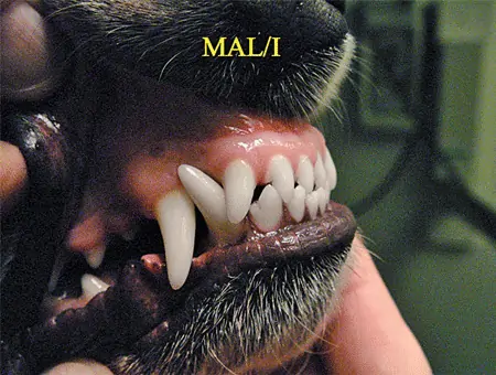
Photo 4: A class 1 malocclusion.
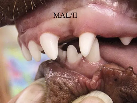
Photo 5: A class 2 malocclusion.
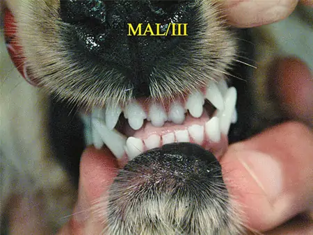
Photo 6: A class 3 malocclusion.
Asymmetrical Malocclusions
According to AVDC, maxillary-mandibular asymmetry describes skeletal malocclusions that can occur in one of the following directions:
Rostrocaudal: A MAL/2 or MAL/3 malocclusion is present on one side of the face while the contralateral side retains normal dental alignment.
Side-to-side: Loss of the midline alignment of the maxilla and mandible.
Dorsoventral: Results in an open bite, defined as an abnormal vertical space between opposing dental arches when the mouth is closed.
Wry bite is a nonspecific layman’s term used to describe various unilateral occlusal abnormalities, so its use isn’t recommended.
Traumatic Malocclusion in a Jack Russell Terrier
This 6 month old Jack Russell Terrier presented for palatal trauma from the mandibular canines. What is the primary problem?
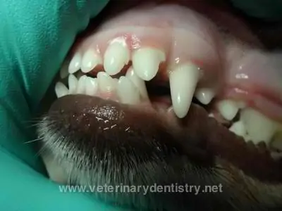
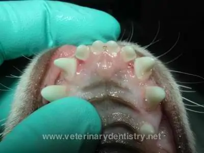
The mandibular canine teeth are linguoverted, resulting in the trauma seen here.
What treatment option would result in the quickest resolution?
Extraction of the second and third incisors bilaterally to allow the mandibular canines to rest adjacent to the extraction site. Due to the significant palatal orientation of the mandibular canines, alveoloplasty and excision of the palatal mucosal margin with a palatally positioned flap was needed to eliminate contact.
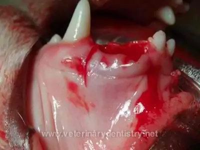
Bilateral vertical releasing incisions were made.
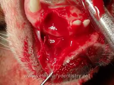
Mucoperiosteum dissected from the underlying musculature, excision of 2 mm of palatal mucosa and alveoplasty performed.
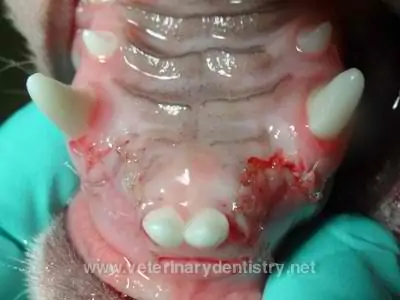
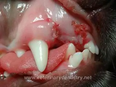
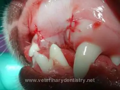
Immediate postoperative images of the palatally positioned flap.
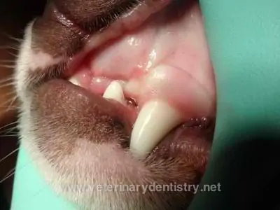
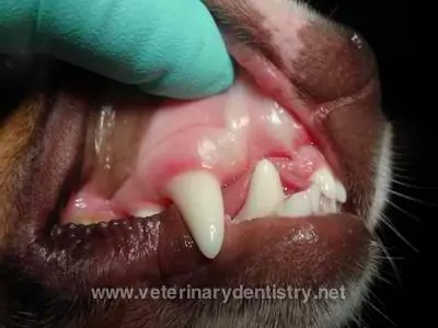
One month postoperative view.
